


 28 / 36

CAMLOG
®
Implant Position Planning
X-RAY TEMPLATE
In the planning template or base produced fromthewax-up/set-up, CT-tubes
for planning or other radio-opaque markers are integrated at the ideal im-
plantation position and are used as reference positions in the x-ray image.
The tubes consist of two parts: the titanium used leaves no scattering on CT
scans. The lower part is polymerized in the template and the upper part in-
serts into this. The complete tube is used in radiologic diagnostics, and the
upper part can be removed during surgery.
The lower part is used during surgery as guiding sleeve for the pilot drill.
Titanium CT-tubes for planning or other radio-opaque positioning compo-
nents (steel, barium sulfate) are integrated, depending on the analysis soft-
ware. If the tubes are placed directly on the mucous membrane, its thick-
ness can be detected on the CT scan.
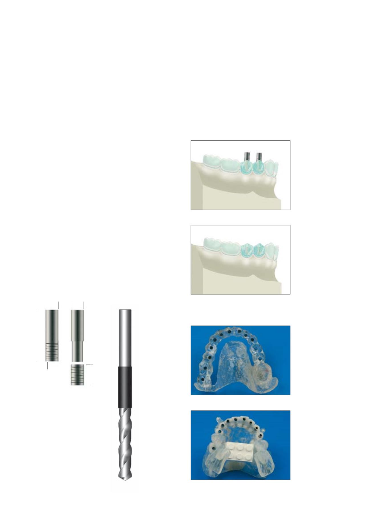
Planning template with CT-tubes
Template without upper sections of tubes for use
as a drilling template
X-ray template, outlined with tubes
X-ray template with radio-opaque teeth and
installed tubes
Ø 2.5 mm external
diameter
10 mm
4 mm
CT-tubes for CT-planning
for drill Ø 2.0 mm,
corrugated tubing, for pilot
drill Ø 2.0 mm
Drill for placement of
CT-tubes, Ø 2.0 mm
Ø 2.1 mm internal
diameter