


 30 / 36

CAMLOG
®
Implant Position Planning
COMPUTER-TOMOGRAPHIC SCAN/DIGITAL
VOLUME TOMOGRAPHY WITH/WITHOUT 3-D
EVALUATION
Precise three-dimensional evaluation of the bone dimensions and subse-
quent planning of the implant positions are only possible using the data
from a CT scan or DVT and a computer-supported planning program. Spe-
cial conditions such as septation or infections in planned sinus floor eleva-
tions and critical vertical relations in the mandible can be recognized. An
x-ray or planning template that the patient wears while the image is taken
provides information about the optimal prosthetic alignment of the im-
plants. Depending on the program, the template must have radio-opaque
position markers (e.g., titanium ball, titanium sleeve, barium sulfate-coated
markers).
With the aid of a CT scan/DVT and a 3-D evaluation, the medical informa-
tion derived can be used to determine the bone quantity and quality (with
DVT, definable only relatively or via calibration). Together with the pros-
thetic information from the geometry of the x-ray template, the number of
implants, implant position, implant diameter and implant length are deter-
mined.
The final prosthetic design and the hard- and soft-tissue augmentation, if
required, are discussed and coordinated in the teamwith the patient on the
basis of this information.
With the information derived, the planned implant positions are checked
and adjusted if necessary.
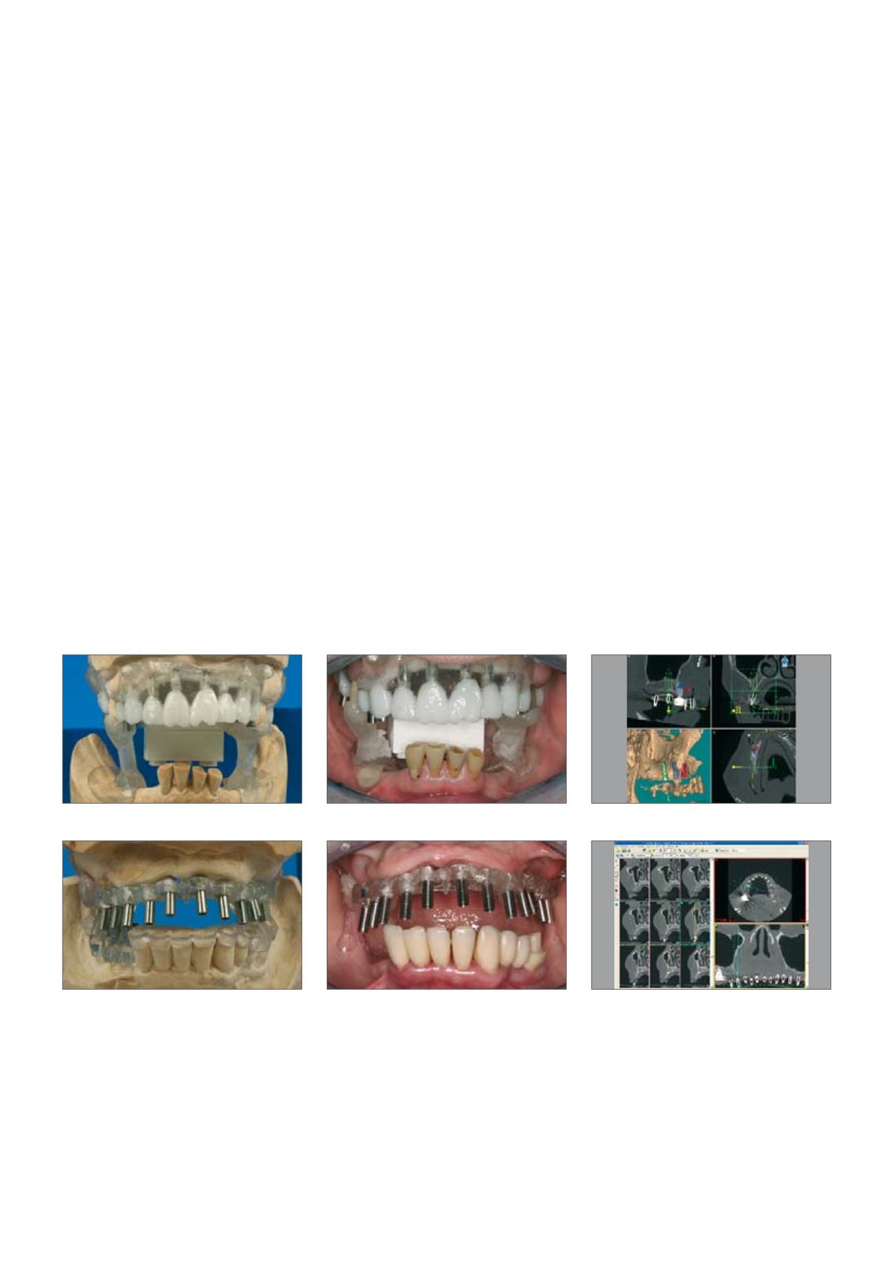
View of the cast positioning
X-ray templates
in situ
Computer-supported 3-D planning
of implant positions
View of the cast positioning
X-ray templates
in situ
Computer-supported 3-D planning
of implant positions